Many CSD students also complete courses in the Honors Program during their time at ACU. These students also complete projects during major’s courses to fulfill Honors College requirements. This year, Ainsley Haley completed a 3-D modeling project of the muscles of the head and neck as an Honors Project in COMP 419 – Speech Science. She has graciously allowed me to post her project on the department blog — I think she did a fabulous job!
Enjoy, Brenda
————————————————————————————————————————————————————————————
Muscles of the Head and Neck
The muscles of the head and neck are complexly related and serve many different functions. I constructed a 3-D model of these muscles and learned a lot about them in the process. I categorized these muscles into four major sections based on their major functions: facial expression, jaw movement, head movement, and swallowing. However, a majority of these muscles have other, over-laid, functions, such as respiration and speech.
I identified ten muscles that where mainly involved in facial expression. The levator labii superioris alaeque nasi muscle dialates the nostril and elevates the upper lip, which results in a snarl. The main function of the levator labii superioris muscle is to elevate the upper lip. The zygomaticus minor muscle draws the upper lip backward, upward, and outward. The zygomaticus major muscle also pulls the angle of the mouth upward and forward, to result in a smile, this function is also shared with the levator anguli oris muscle. Contracting the orbicularis oris closes the mouth or puckers the lips.
The risorius muscle retracts the angle of the mouth, resulting in a smile. The mentalis, also known as the “pouting muscle,” raises and pushes up the lower lip. In contrast, the depressor labii inferioris muscle helps to lower the bottom lip. The depressor anguli oris muscle is associated with frowning, as it depresses the corner of the mouth. The Buccinator muscle pulls back the angle of the mouth and flattens the cheek area. These muscles enable us to exhibit facial expressions, which essential to effective communication. However, some of these muscles also have other vital functions. For instance, the obicularis oris is essential for lip closure during the production of stop phonemes, and the buccinators muscle aids in holding the cheek to the tongue during chewing. The muscles in this group are shown in green in the image below:
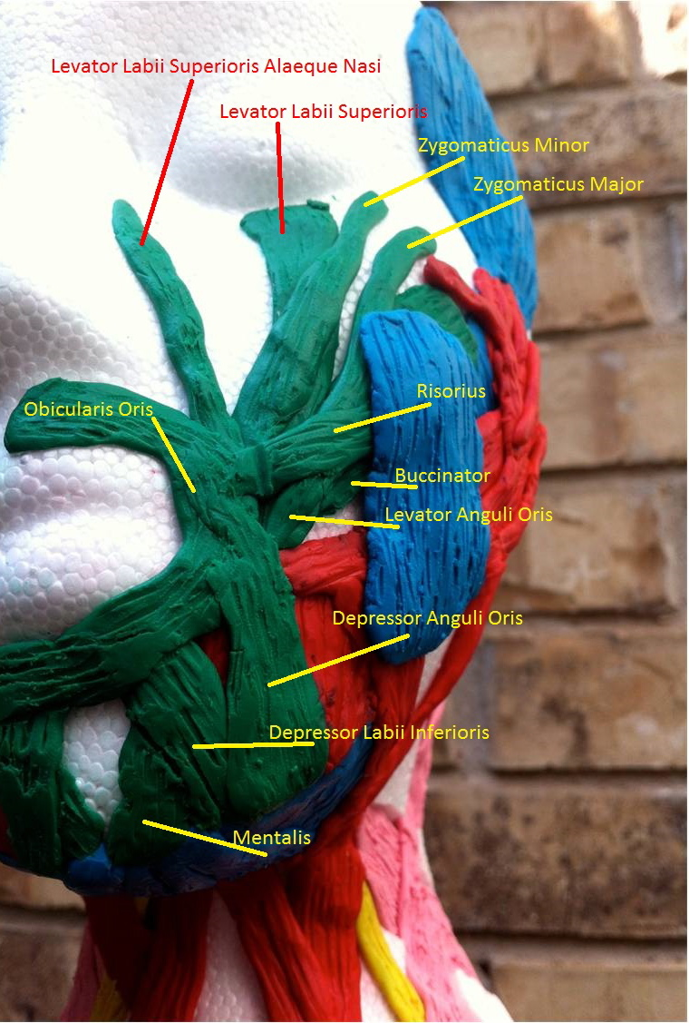
Obicularis oris muscle group
Another major function category is jaw movement. This is essential to both speech and chewing. The temporalis muscle elevates and retracts the mandible. The mylohyoid muscle depresses the mandible, however it also elevates the hyoid, the floor of the oral cavity, and the tongue, which is essential in both swallowing and speaking.
The anterior belly of the digastricus muscle opens the jaw and also draws the hyoid bone forward. The geniohyoid depresses the mandible, and is also active in swallowing. The lateral pterygoid Muscle helps lower the mandible and lower the jaw and also assists in chewing when paired with the medial pterygoid muscle which elevates and closes the jaw. The masseter muscle also works to close the jaw, while the platysma muscle serves to depress the lower jaw. The muscles in this group can be shown in blue in the images below:
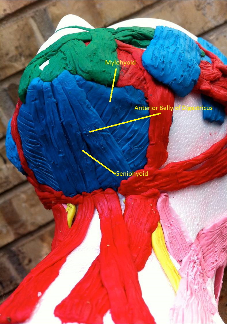
- Jaw muscles – picture 1
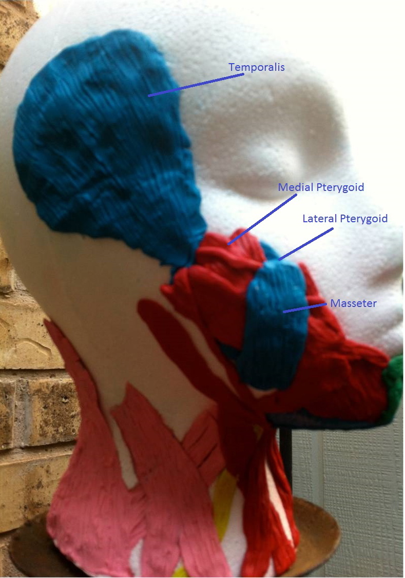
Jaw muscles – picture 2
Jaw movement and swallowing are intricately related. Many of the muscles involved in jaw movement also play a major role in swallowing as well. However, there is a group of muscles whose primary function is swallowing. The superior pharyngeal constrictor muscle is the most superior of the pharyngeal constrictors. It contracts on a bolus when it arrives in the pharynx and propels it down toward the esophagus. The levator veli palatini elevates the soft palate to help prevent food from entering the nasopharynx during swallowing. It is also essential in preventing nasal emission during articulation. Similarly, the tensor veli palatini tenses the soft palate, also to prevent food from entering the nasopharynx during swallowing. The salpingopharyngeus muscle raises the pharynx and the larynx during swallowing and draws the pharyngeal walls up, whereas the sternthyroid depresses the larynx. The stylohyoid draws the hyoid bone backwards and elevates the tongue. The posterior belly of the digastricus elevates the hyoid bone while pulling it posteriorly during swallowing. The sternohyoid depresses the hyoid along with the omohyoid and the thyrohyoid. The palatoglossus elevates the posterior tongue, which aids in the initiation of swallowing. The styloglossus draws up the sides of the tongue to create a trough for swallowing. The hyoglossus depresses and retracts the tongue. The genioglossus is the major muscle responsible for protruding the tongue. These tongue muscles are also important in shaping and forming the tongue into different shapes and placing it in different placements in order to articulate different sounds. The muscles in this group are shown in red in the images below:

Extrinsic tongue muscles – picture 1

Extrinsic tongue muscles – picture 2
The last major function group is head movement. The sternocleidomastoid serves to flex the neck, extend the head, and rotate the head both laterally and contralaterally. The longus capitis muscle flexes the head and neck laterally and rotates the head, as does the splenius capitis muscle. The trapezius, levator scapulae muscle, and scalene muscles all serve to move the head, however they all play an important role in respiration as well. The muscles that are involved in head movements are shown in yellow and pink in the images below, the muscles in pink are those that also function as muscles of respiration:

Muscles controlling head movement – picture 1
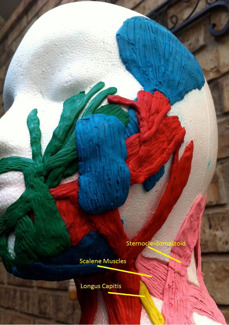
Muscles controlling head movement – picture 2