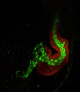
Confocal image of a developing zebrafish heart
Transgenic line (myl7GFP:flkmcherry) allowing us to visualize heart (green) and vascular (red) tissues
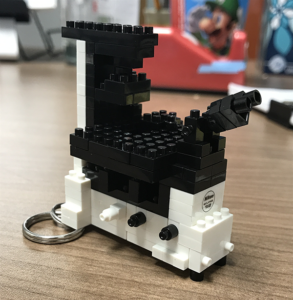
Lego model of a Nikon eclipse Ti2 confocal microscope
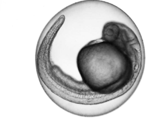
Developing zebrafish larvae at 26 hours post fertilization
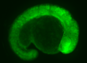
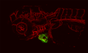
Zebrafish embryo injected with GFP mRNA
Holowiecki lab leather notebook made at the ACU Maker Lab